Concomitant Pulmonary Hamartoma and Azygos Lobe
1Department of Emergency Medicine, Amasya University Sabuncuoglu Serefeddin Research and Training Hospital, Amasya, Turkey; 2Department of Pulmonology, Kars Harakani State Hospital, Kars, Turkey
![]() 10.3329/bmrcb.v45i2.42547
10.3329/bmrcb.v45i2.42547 ![]() 0000-0003-2727-0556
0000-0003-2727-0556
Lungs are located either sidess of heart in thoracic cavity. The left lung is seperated by one fissure and right lung is seperated by oblique and horizontal fissures.1 0.4%-1% of anatomical human dissections showed that azygos lobe is an accessory lobe due to a defect of the migration of vena azygos during the intrauterine development. Normally, azygos vein cross over the apex of the right lung and reaching to superior vena cava. In rare cases, azygos vein penetrates & through the upper lobe of the right lung and reaches the pleura and it consists of an accessory fissure called with "azygos fissure". This variational structure usually cause any symptoms and it is found by incidentally.2
Pulmonary hamartoma is the most common benign tumor of the lung and this lesion has a male predominance. These lesions are usually diagnosed incidentally on chest radiographs.3
Hamartomas can be located parenchymal or endobronchial.4 Intraparenchymal hamartomas generally do not cause symptoms but endobronchial lesions may cause cough, fever and pulmonary infection due to obstruction. 5 Herein, it was aimed to present a case of concomitant pulmonary hamartoma and azygos lobe.
A 50 year woman admitted to hospital complaining with dry cough that began 3 week before admission to the hospital. Physical examination revealed no pathological finding. The patient’s medical history was unremarkable. The patient did not have a smoking history and there was no abnormal value in the laboratory findings.
On her chest radiograph,the patient had a superposed 1.5 cm coin lesion at right paratracheal area which was found in a variant azygos lobe in right superior lobe (figure 1).
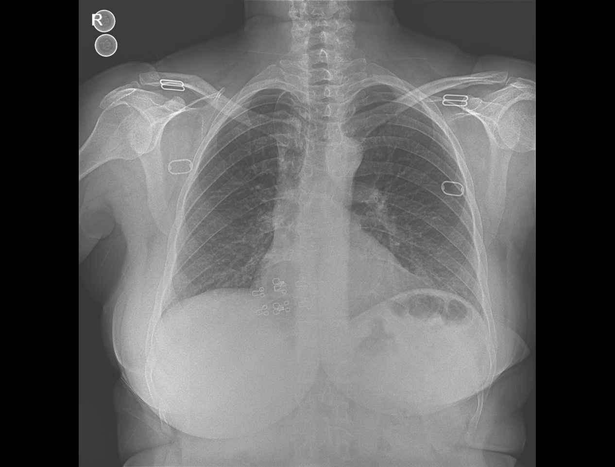
Figure 1:First Admission Chest X-Ray
Thoracic tomography was performed because of the lesion detected on chest radiograph.On thoracic computed tomography, the patient had a solid mass in the azygos lobe of 15x14 mm in the right lung with an average density of -10 HU (fat density).This lesion was thought to be a pulmonary hamartoma. The patient was in follow-up for a year and no change was observed radiologically (figure 3).
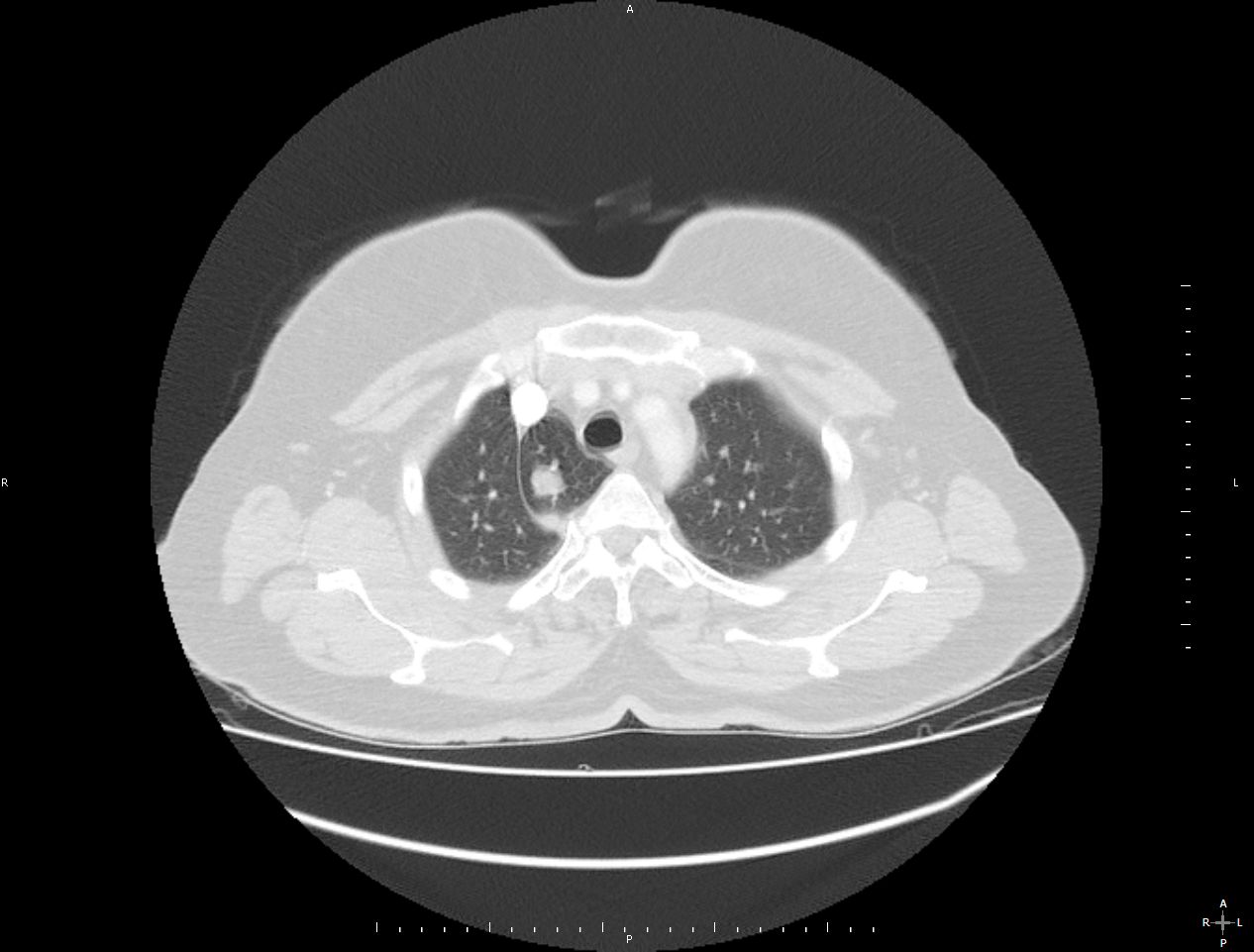
Figure 2:First Admission Thorax CT
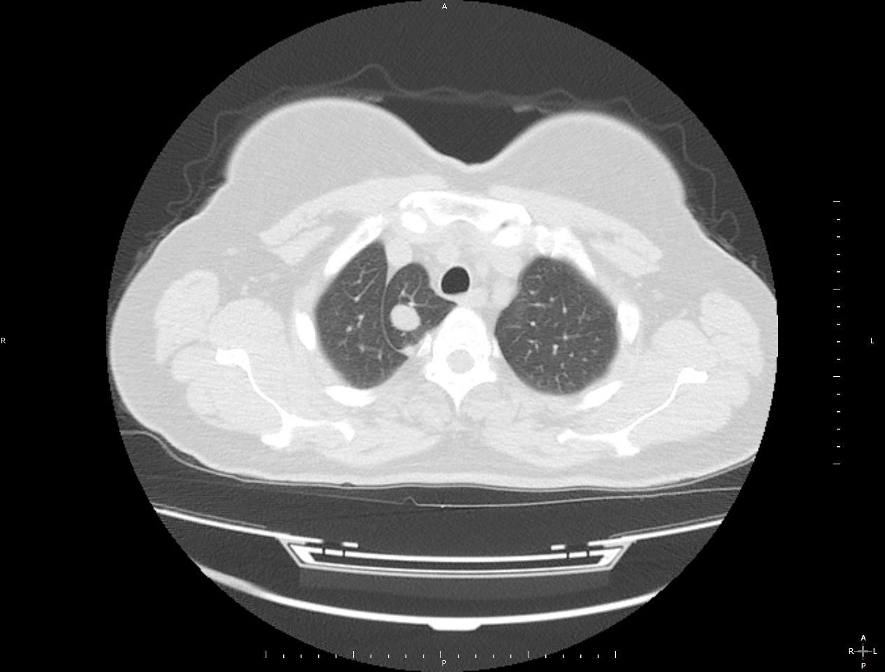
Figure 3: Control Thorax CT One Year Latesr
Pulmonary hamartoma is the most common benign lung lesion and causes symptoms when it reaches large size or endobronchial location. The azygos lobe is a rare anatomical variation and it is usually found in the right lung. In this patient, there were no symptoms of these diagnoses. Sometimes, azygos lobe can be misdiagnosed as an enlarged thymus, a substernal guatr, a small sized pneumothorax or neoplasm. Azygos lobe is a benign and common variation anomaly of the lung but its behaviour in pathological conditions is very important. The short letter was aimed to present a rare radiological togetherness of azygos lobe and pulmonary hamartoma.
References
- Sudikshya KC, Shrestha P, Shah AK, Jha AK, Variations in Human Pulmonary Fissures and Lobes: a Study Conducted in Nepalese Cadavers. Anat Cell Biol 2018;51:85-92
- Kotov G, Dimitrova IN, Iliev A, et al. (June 11, 2018) A Rare Case of an Azygos Lobe in the Right Lung of a 40-year-old Male. Cureus 10(6): e2780.
- Acar M, Ozates M, Ekici F,SimSek M. Tanısal ve Girisimsel Radyoloji (2001) 7:373-375
- Geramizadeh, B, Mottavvas M, Zeyaian B & Amirian, A. (2019) Giant Hamartoma of Lung Presented with Massive Hemoptysis: A Rare Case Report and Review of the Literature. Rare Tumors. 2019; 11: 2036361318823926.
- Evkan A, Komurcuoglu B, Kaplan G, Guvencli M, Yalnız E, Salik B, Izmir Gögüs Hastanesi Dergisi, Cilt XXX Sayı 1, 2016